
Why BMI Is Inaccurate: The Evidence-Based Guide
20 August 2026Updated 25 August 2026
Body mass index, or BMI, is a useful screening measure, but it is not a direct measurement of body fat or a complete assessment of health. It combines weight and height, so it cannot distinguish fat from lean tissue, show where fat is stored or identify obesity-related organ dysfunction.
Table of Contents
- What BMI Measures
- Where BMI Can Be Inaccurate
- Ethnicity and Current UK Thresholds
- Waist and Central Adiposity
- Body Composition and DEXA
- A More Complete Health Assessment
What BMI Measures
BMI is calculated as weight in kilograms divided by height in metres squared:
BMI = weight (kg) ÷ height² (m²)
For most White adults, standard UK categories define 18.5–24.9 kg/m² as the healthy-weight range, 25–29.9 as overweight and 30 or above as obesity. BMI below 18.5 can also be clinically important and may prompt assessment for undernutrition or another cause of low weight.
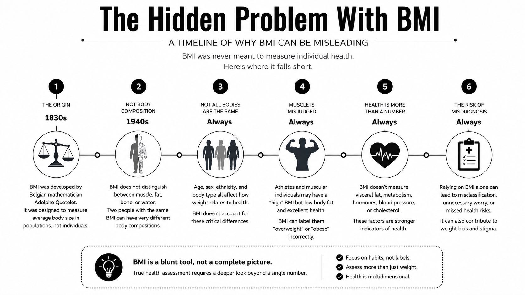
Belgian statistician Adolphe Quetelet described the weight-to-height-squared relationship in the 1830s while studying populations. The term body mass index and its modern epidemiological use developed later, including work by Ancel Keys and colleagues in 1972. This Lancaster University report on BMI history discusses the social and clinical development of the measure.
Its origin does not make BMI automatically invalid. BMI is inexpensive, reproducible and associated with health outcomes at population level. It is also useful in primary-care screening. The error arises when the category is treated as a direct diagnosis of excess fat or a complete account of one person’s health.
Where BMI Can Be Inaccurate
BMI cannot separate fat mass, lean soft tissue, bone mineral or fluid. This creates several recognised limitations:
- High muscle mass: a muscular person may have an elevated BMI without excess adiposity.
- Ageing: loss of muscle and height can change the meaning of a stable BMI.
- Fluid changes: pregnancy, oedema and some medical conditions alter weight without reflecting fat gain.
- Fat distribution: two people with the same BMI can have different amounts of abdominal fat.
- Ethnicity: cardiometabolic risk can occur at lower BMI in some populations.
Practical rule: BMI is a screening data point, not a standalone diagnosis or verdict.
An online calculator such as this commercial XO BMI tool can perform the arithmetic. Its result still requires the same clinical context and appropriate ethnicity-specific thresholds.
Ethnicity and Current UK Thresholds
NICE advises lower thresholds for adults from South Asian, Chinese, other Asian, Middle Eastern, Black African or African-Caribbean backgrounds because central adiposity and cardiometabolic risk occur at lower BMI on average:
- Overweight: BMI 23–27.4 kg/m²
- Obesity: BMI 27.5 kg/m² or above
These are group-level practical thresholds, not proof that every person within an ethnic category has the same body composition or risk.
A 2025 Guardian report covered Nesta modelling that applied lower thresholds to English survey data. It estimated that official statistics understated overweight and obesity prevalence among minority ethnic adults. The “one million” estimate was a modelling result, not direct examination showing that one million individuals had each received an incorrect clinical diagnosis.
The current NICE guidance on identifying overweight, obesity and central adiposity is the appropriate source for clinical thresholds.
Waist and Central Adiposity
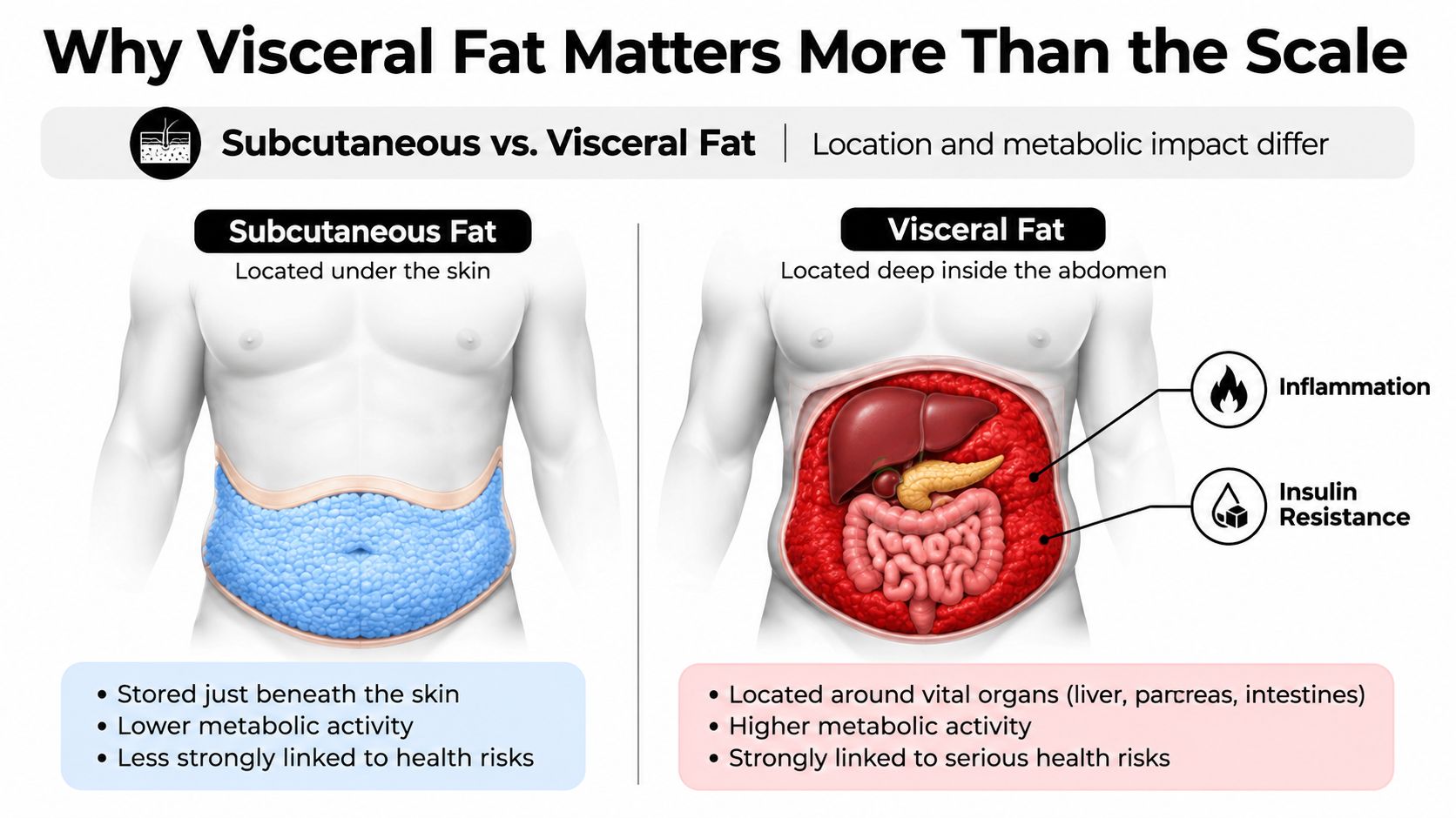
Central adiposity refers to fat stored around the abdomen. It is associated with increased risk of type 2 diabetes, hypertension and cardiovascular disease. BMI does not show fat distribution, so a waist measure can add useful information.
For adults with BMI below 35, NICE classifies waist-to-height ratio as:
- 0.4–0.49: healthy central adiposity range;
- 0.5–0.59: increased central adiposity and health risk;
- 0.6 or above: high central adiposity and further increased risk.
The simple public message is to keep waist circumference below half of height. Waist-to-height ratio is still a screening measure, not a direct visceral-fat scan. Technique, pregnancy and some medical conditions can affect interpretation.
The House of Lords Library briefing on BMI summarises UK debate about BMI, muscularity and central adiposity. It is a policy briefing rather than a clinical guideline.
Body Composition and DEXA
A hypothetical example shows the limitation. Two adults who are both 1.80 m tall and 88 kg each have a BMI of 27.2. One may have high muscle mass and a small waist; the other may have less muscle and more abdominal fat. BMI cannot distinguish them.
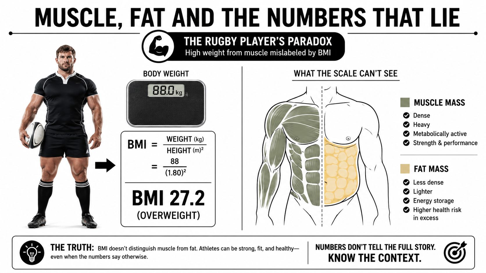
A DEXA body-composition scan estimates total and regional fat mass, lean soft tissue and bone mineral density. It does not directly measure skeletal muscle, and its estimate of visceral adipose tissue is not equivalent to direct CT or MRI measurement.
DEXA is a useful reference method but not an infallible “gold standard”. Hydration, positioning, device software and biological variation can affect results. The scan also uses a small dose of ionising radiation and is not necessary for routine BMI screening.
A More Complete Health Assessment
| Question | Useful information |
|---|---|
| Is weight high or low relative to height? | BMI with appropriate thresholds |
| Is fat concentrated centrally? | Waist circumference or waist-to-height ratio |
| What are fat, lean-soft-tissue and bone estimates? | DEXA or another validated body-composition method where indicated |
| Is cardiometabolic risk elevated? | Blood pressure, medical history and clinician-selected blood tests |
| Is physical function affected? | Strength, mobility, symptoms and daily function |
No single test answers every question. Bioelectrical impedance is convenient but affected by hydration and device equations. Skinfold assessment depends strongly on assessor skill and estimates subcutaneous fat. CT and MRI can characterise abdominal fat more directly but are not routine screening tools.
NICE also recommends caution when interpreting BMI in adults with high muscle mass and in people aged 65 or older. In later life, comorbidity, functional capacity, unintentional weight loss and the possible protective effect of a slightly higher BMI all influence interpretation.
Seek clinical assessment for unexplained weight change, a rapidly changing waist, swelling, persistent fatigue or other concerning symptoms. A clinician can decide whether blood pressure, HbA1c, glucose, lipids, thyroid testing or other investigations are appropriate. Tests such as fasting insulin or CRP should not be ordered or interpreted routinely without a clinical reason.
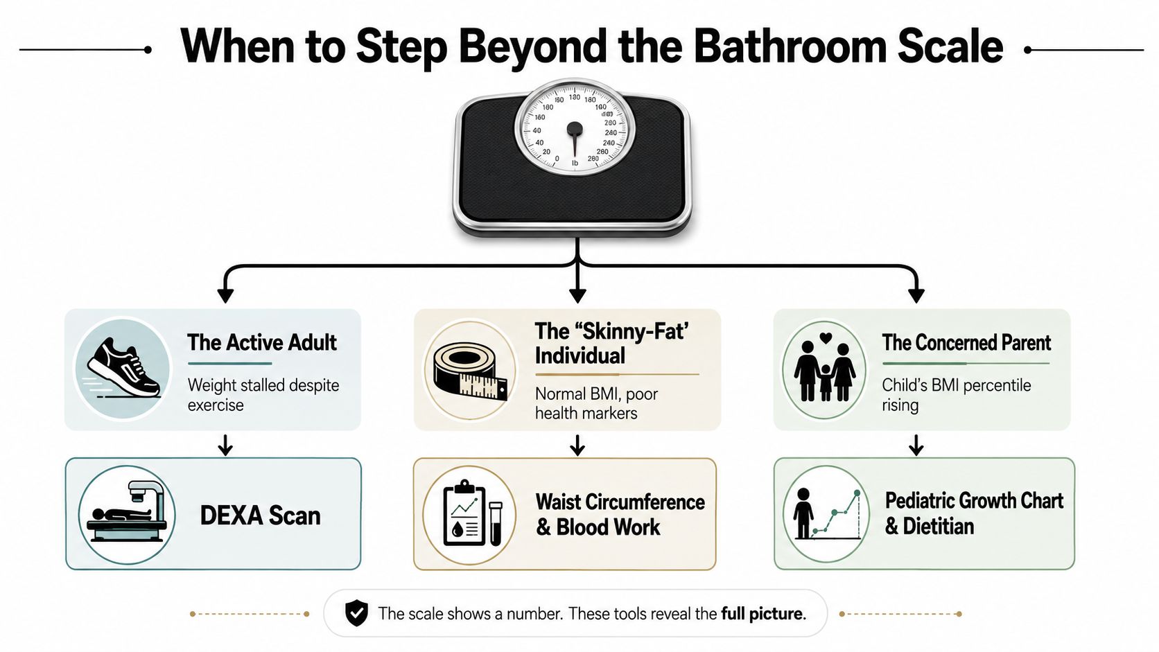
BMI should therefore be demoted from a verdict to one part of a wider assessment. It remains useful because it is simple and evidence-linked, while waist measures, body composition, function and clinical markers supply information the formula cannot.
The content in this article is for educational purposes only and does not constitute medical or dietary advice. If you have an underlying health condition, are taking medication, or are considering significant changes to your diet or exercise regimen, consult a qualified healthcare professional before making any adjustments.